
Everyday Habits That Can Protect Your Eyes from Damage
4 Min Read

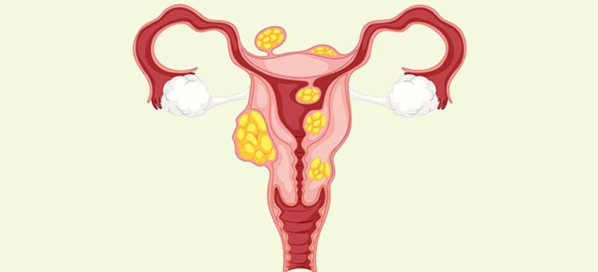
Uterine fibroids, also known as leiomyomas, are non-cancerous growths that develop in the uterus. These common muscular tumors affect many women, particularly during their reproductive years. Advancements in interventional radiology techniques have opened up non-surgical options that provide effective alternatives. This article will explore various interventional radiology techniques for treating uterine fibroids.
Uterine fibroids are benign tumors that appear in the muscular wall of the uterus. They can vary in size, ranging from small seedlings to large masses. These masses can distort the shape of the uterus. Although the exact cause of fibroid formation is unknown, hormonal imbalances and genetic factors could play a role. According to an interventional radiologist in El Paso, uterine fibroids can cause a various symptoms, including:
If a woman experiences the symptoms of uterine fibroids, a doctor will perform a thorough physical examination. They may recommend more tests, such as an hysteroscopy, to confirm the diagnosis.
Historically, the treatment of uterine fibroids has primarily involved surgical interventions. Medications such as hormonal therapies can help manage symptoms. However, they do not eliminate the fibroids. While these surgical procedures can be effective, they are invasive and may involve longer recovery.
Interventional radiology techniques offer non-surgical alternatives for the treatment of uterine fibroids.
Interventional radiologists perform these minimally invasive procedures. They use image guidance to target and treat the fibroids precisely. Here are some commonly used non-surgical interventional radiology techniques for uterine fibroid:
UFE is a procedure that involves blocking the blood supply to the fibroids. During the procedure, tiny particles are injected into the uterine arteries. These particles cut off the blood flow to the fibroids, causing them to shrink and eventually disappear. UFE is performed under local anesthesia, and most patients can return home the same or the following day.
MRgFUS uses focused ultrasound waves to heat and destroy the fibroid tissue. Real-time MRI images guide the procedure. This technique allows to target the fibroids while sparing the surrounding healthy tissue. During the procedure, patients can experience minimal discomfort.
RFA utilizes heat energy to destroy the fibroid tissue. During the procedure, a thin needle-like probe is inserted into the fibroid. Then radiofrequency energy is transmitted to heat and ablate the fibroid cells. RFA can be performed under local anesthesia, and patients can experience minimal pain.
Cryomyolysis is a technique that involves freezing the fibroid tissue. During the procedure, a special probe is inserted into the fibroid. Extremely cold temperatures are applied to freeze and destroy the fibroid cells. Cryomyolysis requires local anesthesia, and patients may experience mild cramping.
HIFU uses focused ultrasound waves to heat and destroy the fibroid tissue. The ultrasound waves target the fibroids, causing tissue necrosis.
Non-surgical interventional radiology techniques offer several advantages over surgical treatments for uterine fibroids. Some of the key benefits include:
Non-surgical interventional radiology techniques have revolutionized the treatment landscape for uterine fibroids. These minimally invasive procedures are highly effective and enable faster recovery times. With advancements in interventional radiology, women now have many choices when managing their uterine fibroids.

What Does Black Lines On Nails Mean?
9 Min Read

9 Effective Uses of Expired Olive Oil
7 Min Read

Disadvantages Of Drinking Lemon Water Daily
7 Min Read

List Of Vitamins That Cause Weight Gain
5 Min Read
How To Get Rid Of Blood Clot In Finger?
8 Min Read
Disclaimer - The content on this site is for informational purposes only. Always seek the advice of a qualified physician.
Copyright © 2025 sehat.com